Why is Anterior Segment Ocular Photography Important?
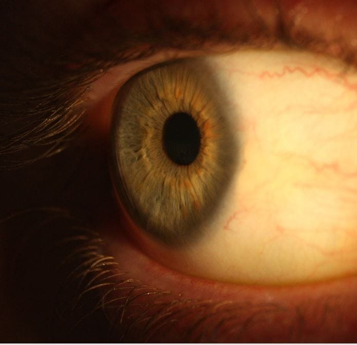
Anterior segment ocular photography involves taking detailed digital photographs of the front parts of your eye. This includes the cornea (the clear front surface), iris (the colored part), pupil (the black center), and lens.
These photographs provide your eye care professional with a valuable and permanent record of the appearance of the front of your eye at a specific point in time. This is important for several reasons:
- Documentation and Baseline: It establishes a baseline view of your eye’s health, allowing for comparison during future examinations to detect even subtle changes.
- Monitoring Progression: For existing eye conditions, photography helps track their progression or response to treatment over time with objective visual evidence.
- Enhanced Communication: The photographs can be used to explain your condition more clearly to you and, if necessary, to other healthcare professionals involved in your care.
- Detecting Subtle Abnormalities: High-resolution images can reveal minute details that might be difficult to fully document with a written description alone.
What Conditions Can Be Managed with Anterior Segment Ocular Photography?
Anterior segment photography is a useful tool in managing a wide range of eye conditions, including:
- Corneal Issues: This includes conditions like corneal abrasions, ulcers, infections (e.g., keratitis), scars, and irregularities. Photography helps document their size, location, and severity.
- Conjunctival Issues: Problems affecting the conjunctiva (the clear outer layer of the white of your eye and the inside of your eyelids), such as infections (conjunctivitis), growths (e.g., pinguecula, pterygium), and vascular abnormalities, can be effectively documented.
- Iris and Pupil Abnormalities: Changes in the shape, size, or color of the iris, as well as unusual pupil reactions or irregularities, can be captured and monitored.
- Cataracts: While photography doesn’t fully assess the density of a cataract, it can document its initial appearance and progression over time.
- External Eye Diseases: Conditions affecting the eyelids and surrounding structures can sometimes be documented to provide a comprehensive record.
- Contact Lens Complications: Photography can help document any adverse effects of contact lens wear on the front of the eye.
- Post-Surgical Monitoring: Following eye surgery, photographs can help track the healing process and identify any potential complications.
What to Expect During Anterior Segment Ocular Photography?
The anterior segment photography procedure is quick, painless, and non-invasive. Here’s what you can typically expect at our Dalby practice:
- Positioning: You will be asked to sit comfortably in front of a specialized camera, usually mounted on a slit lamp (the same instrument used for routine eye examinations). Your chin will rest on a chin rest, and your forehead will be against a forehead strap to help keep your head still.
- Focusing: Your eye care professional will carefully focus the camera on the front part of your eye using the slit lamp’s magnification.
- Multiple Images: Depending on the reason for the photography, several images may be taken of different parts of the front of your eye or from different angles.
- Duration: The entire process usually takes just a few minutes.
- No Discomfort: You will not feel any pain or discomfort during the procedure. There are no eye drops required specifically for the photography itself.
The digital images captured will be securely stored in your patient record and can be reviewed by your eye care professional during your consultation. If you have any questions about anterior segment ocular photography, please feel free to ask our friendly team here in Dalby! We are committed to providing you with the best possible eye care.
Meet Our Optometrists: Tom Roger & Tobin Eapen
The Eyecare Eyewear Optometry Team has been trained to provided you with comprehensive, personalised advice regarding your current and future visual needs. The combination of years of experience and an accomplished, professional optical dispensing team means that you will receive the highest level in vision care and ocular disease management.